Unveiling a New Dimension in Dental Care: The Era of Dental CBCT Machines
In the ever-evolving landscape of modern dentistry, the introduction of dental Cone Beam Computed Tomography (CBCT) machines marks a revolutionary leap. These advanced imaging systems have ushered in a new era, transcending the limitations of traditional two-dimensional radiography and opening up a world of three-dimensional diagnostic possibilities. Today, dental CBCT machines stand at the forefront of dental technology, offering unparalleled insights into oral anatomy and pathology.
The significance of dental CBCT in contemporary dental practice cannot be overstated. By providing detailed 3D images of a patient’s teeth, jaw, and surrounding structures, CBCT Machines have transformed the way dental professionals approach diagnosis and treatment planning. This leap from flat images to volumetric visualization equips dentists with a depth of understanding previously unattainable, paving the way for more precise, efficient, and personalized dental care.
As we delve into the world of 3D dental imaging, it’s essential to recognize the profound impact CBCT technology has had on various dental specialties. From intricate implant placements and endodontic assessments to complex orthodontic evaluations and oral surgery planning, the applications of CBCT are as diverse as they are transformative.
In this blog, we will explore the journey of dental CBCT machines, examining how they have redefined dental diagnostics and treatment. We will also look ahead to what the future holds for this groundbreaking technology and its potential to further revolutionize the field of dentistry, promising enhanced patient care and more accurate clinical outcomes.
The Evolution of Dental Imaging Technology
From X-Rays to 3D Visualization
The journey of dental imaging technology is a tale of continuous innovation and transformative breakthroughs. From the early days of conventional X-ray imaging, which provided the first glimpses beneath the oral surface, dental imaging has evolved dramatically. Traditional two-dimensional X-rays, while revolutionary at their inception, had limitations in terms of detail and perspective, often leaving crucial aspects of dental anatomy to the imagination.
The advent of Cone Beam Computed Tomography (CBCT) marked a pivotal moment in this evolutionary timeline. CBCT emerged as a game-changer, offering a three-dimensional view of the oral and maxillofacial region. This technology uses a cone-shaped X-ray beam that rotates around the patient, capturing a multitude of images from different angles. These images are then reconstructed into a 3D model, providing a comprehensive view of the patient’s dental structures.
Transformative Impact of CBCT in Dental Diagnostics
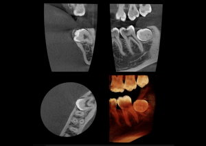
The introduction of CBCT has revolutionized dental diagnostics by bringing unprecedented precision and depth to imaging. Unlike traditional X-rays, CBCT provides detailed visualization of bone structure, nerve pathways, soft tissues, and even airways in three dimensions. This level of detail is critical in diagnosing complex cases, planning surgical procedures, and formulating more effective treatment strategies.
CBCT’s impact is particularly significant in areas requiring high precision, such as implantology, endodontics, and orthodontics. In implant dentistry, for example, CBCT aids in accurate implant placement, considering the bone density and the proximity to vital structures. In endodontics, it allows for better visualization of root canal anatomy, leading to more successful root treatments.
Furthermore, the depth of information provided by CBCT assists in identifying potential issues that may not be apparent in 2D imaging, such as hidden infections or tumors. This comprehensive diagnostic capability not only enhances treatment accuracy but also significantly improves patient safety.
The Mechanics of CBCT: Understanding 3D Imaging
Unraveling the Technology Behind CBCT
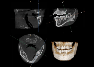
Cone Beam Computed Tomography (CBCT) represents a significant advancement in dental imaging technology, fundamentally differing from standard radiography in both its mechanics and output. At the heart of CBCT technology is a cone-shaped X-ray beam, which is the key to its ability to produce three-dimensional images. Unlike traditional radiography that captures flat, two-dimensional images, CBCT rotates around the patient, capturing data from multiple angles using this cone-shaped beam.
This data is then reconstructed digitally to create a detailed three-dimensional representation of the patient’s dental structures. The process involves sophisticated algorithms that process the X-rays captured from different angles, compiling them into a single 3D image. This image provides a comprehensive view of the teeth, jaws, bone structure, and even soft tissues and nerves, which is not achievable with standard 2D X-rays.
Benefits of 3D Dental Imaging
The transition from 2D to 3D imaging offers numerous benefits, particularly in terms of precision and clarity. One of the primary advantages of CBCT is its ability to provide a more accurate and detailed view of dental anatomy. This precision is crucial for various aspects of dental care, including diagnosis, treatment planning, and surgical execution.
In terms of clarity, CBCT images offer unparalleled resolution, allowing dentists to identify and evaluate even the minutest structures with great accuracy. This level of detail is particularly beneficial for complex diagnoses and procedures, such as identifying the exact location of impacted teeth, assessing bone quality for implants, or planning orthodontic treatments.
Furthermore, the precision and clarity of CBCT imaging significantly reduce the risk of unexpected complications during procedures. By providing a complete and detailed view of the area of interest, CBCT allows dentists to plan and execute treatments with a higher degree of confidence and safety.
Innovations in Dental Radiography
Recent Advancements in CBCT Technology
The field of dental radiography has witnessed remarkable advancements in recent years, particularly with the development of Cone Beam Computed Tomography (CBCT) technology. These innovations have not only refined the imaging process but have also expanded the capabilities of dental diagnostics, allowing for more detailed and accurate assessments.
One of the key advancements in CBCT technology is the significant improvement in image resolution and quality. Modern CBCT machines are capable of producing high-resolution images that offer unprecedented clarity and detail. This advancement is crucial for accurate diagnosis and treatment planning, especially in complex dental cases where precision is paramount.
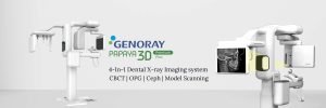
Genoray Papaya 3D Plus: A Case in Point
The Genoray Papaya 3D Plus represents a significant leap in dental digital imaging technology. This cutting-edge Cone Beam Computed Tomography (CBCT) device is a testament to Genoray’s commitment to revolutionizing dental diagnostics. It seamlessly integrates 3D CT, Panoramic, and optional Cephalometric imaging, catering to a wide array of diagnostic requirements. Its versatile imaging capabilities are crucial for precise implant planning and other dental procedures.
Key features of the Genoray Papaya 3D Plus include:
– Multi-Modal Imaging: It combines 3D CT, panoramic, and optional cephalometric imaging, ensuring comprehensive diagnostic information.
– Award-Winning Design: Inspired by the smooth contours of a pebble, it has been recognized for its design by the Korea Ministry of Design Motivation and KEIT, showcasing its innovative R&D convergence.
– Dedicated Sensors: It boasts three distinct CMOS flat panel sensors, each tailored for specific imaging needs, ensuring durability and optimal performance without overloading a single sensor.
– Scout Mode: This feature helps in minimizing positioning errors, enhancing the accuracy of scans.
– Smart Metal Artifact Removal (SMARF): This technology significantly improves image clarity by removing artifacts.
– Rapid Cephalometric Imaging: The 2Sec Ceph (plus) technology captures cephalometric images in just 2 seconds, reducing the chances of patient movement and enhancing image quality.
– Versatile Scan Modes: It offers three scan modes – Normal, Fast, and HD, along with 19 Field of View (FOV) options to suit various dental practices.
– Specialized FOV for Different Cases: This includes 45 cm FOV with 75-micron voxel for Endodontic cases, up to 1614 cm FOV with 200-micron voxel for Maxillofacial cases.
– Efficient Scanning and Reconstruction: Scanning times range from 7.7 to 14.5 seconds, with less than a minute required for image reconstruction.
– Model Scanning Capability: The Papaya 3D Plus can scan both impressions and plaster casts. Utilizing the advanced THEIA software, it provides immediate digital STL format models, reducing the need for physical plaster casts.
– Image Sharing and Compatibility: Images are stored in DICOM® format, supporting open software architecture and allowing for flexibility in software solutions.
– Robust and Accessible Design: The sealed base and columns protect against pests, and its self-standing, wheelchair-accessible design negates the need for wall or floor drilling.
Impact on Patient Outcomes
The innovations in CBCT technology, as exemplified by the Genoray Papaya 3D Plus, have a direct and significant impact on patient outcomes. High-resolution dental scans provide dentists with a comprehensive view of the dental structures, enabling them to detect issues that may not be visible with traditional imaging methods. This level of detail aids in early diagnosis and more accurate treatment planning, reducing the likelihood of complications and the need for invasive procedures.
Moreover, the enhanced imaging capabilities of modern CBCT machines like the Genoray Papaya 3D Plus allow for more precise surgical planning, particularly in implantology. This precision ensures better alignment and placement of implants, leading to improved functional and aesthetic outcomes for patients.
CBCT Applications in Dentistry
Diverse Uses Across Dental Specialties
Cone Beam Computed Tomography (CBCT) has revolutionized dental practice with its versatility and comprehensive imaging capabilities. Its applications span across various dental fields, each benefiting uniquely from the detailed three-dimensional insights provided by this technology.
In Orthodontics: CBCT is invaluable for orthodontic assessment and treatment planning. It allows for precise measurements of craniofacial structures, aiding in the diagnosis of malocclusions and planning of orthodontic interventions. With CBCT, orthodontists can visualize the exact positioning of teeth, including impacted ones, and assess the relationship between teeth and surrounding bone structures, which is crucial for effective brace placement or aligner therapy.
In Oral and Maxillofacial Surgery: Oral surgeons leverage CBCT for pre-surgical planning and navigation. The technology is particularly useful for complex extractions, dental implant placements, and reconstructive surgeries. CBCT provides detailed images of bone structure, nerve pathways, and soft tissues, enabling surgeons to plan procedures that minimize the risk to critical structures like the inferior alveolar nerve.
In Endodontics: Endodontists use CBCT for diagnosing and treating complex root canal cases. The technology aids in identifying root canal anomalies, such as extra canals or unusual anatomy, and in assessing root fractures and the extent of periapical infections. This detailed visualization ensures more precise root canal treatments and better outcomes.
In Periodontics: In periodontal treatment, CBCT assists in assessing bone loss, planning regenerative procedures, and placing dental implants in patients with periodontal disease. It offers a clear view of bone quality and quantity, which is essential for successful periodontal therapies.
In Implant Dentistry: CBCT has become a standard in implantology for its ability to aid in precise implant planning. It allows dentists to assess bone density, determine the optimal location for implant placement, and avoid vital structures such as nerves and sinuses. This precision significantly increases the success rate of implant surgeries.
Contributions to Advanced Diagnostics and Planning
The applications of CBCT extend beyond mere visualization; they contribute significantly to advanced dental diagnostics and treatment planning. By providing a complete and accurate view of dental and facial anatomy, CBCT allows dentists to diagnose conditions that may be missed with traditional imaging methods. This comprehensive approach leads to more informed decision-making, tailored treatment plans, and ultimately, improved patient care.
CBCT’s role in minimally invasive surgery planning also deserves mention. By allowing detailed preoperative assessment, CBCT minimizes surgical explorations, reduces patient discomfort, and speeds up recovery times.
The Future of Dental Imaging
Anticipating Developments in CBCT Technology
As we look to the future of dental imaging, it is clear that Cone Beam Computed Tomography (CBCT) technology will continue to be at the forefront of innovation and advancement. The potential for future developments in this field is vast and holds promise for even more sophisticated and efficient diagnostic capabilities.
One of the key areas of future development in dental CBCT technology is likely to be the enhancement of image resolution and clarity. As imaging technology evolves, we can expect CBCT machines to produce even more detailed and higher-resolution images, allowing for an even greater level of precision in diagnosis and treatment planning.
Another anticipated advancement is the integration of artificial intelligence (AI) and machine learning into dental imaging. AI could be used to analyze CBCT scans more efficiently and accurately, identifying potential issues more quickly and assisting in creating more effective treatment plans. Machine learning algorithms could also help in the automatic detection of common dental conditions, streamlining the diagnostic process.
Potential Advancements and Implications
The future of dental CBCT technology may also include more streamlined and compact designs, making these systems more accessible to a wider range of dental practices, including smaller clinics. This would democratize high-quality imaging technology, making advanced diagnostics available to a larger patient population.
Furthermore, advancements in 3D printing and digital dentistry could be closely tied with CBCT technology. High-resolution CBCT scans could be used in conjunction with 3D printing for creating precise dental restorations, orthodontic devices, and even surgical guides, further enhancing the accuracy and efficiency of dental treatments.
Another exciting possibility is the development of real-time imaging capabilities. This innovation would allow dentists to view CBCT scans in real-time during procedures, providing invaluable guidance during complex surgical interventions.
The implications of these advancements for dental care and diagnostics are significant. With more accurate and detailed imaging, dental professionals can diagnose conditions earlier and more accurately, leading to better patient outcomes. The efficiency gained through these technologies also means that patients can enjoy quicker and less invasive procedures, improving the overall dental care experience.
Conclusion
Reflecting on the Evolution of Dental CBCT Machines
As we reach the conclusion of our exploration into the world of dental CBCT machines, it’s evident that this technology has embarked on an extraordinary journey, significantly shaping the landscape of modern dentistry. From their inception as a novel approach to dental imaging to their current status as indispensable tools in a myriad of dental specialties, CBCT machines have continuously pushed the boundaries of diagnostic precision and efficiency.
The current state of dental CBCT technology reflects a harmonious blend of advanced imaging capabilities, patient-centered design, and practical application in clinical settings. These machines have revolutionized dental diagnostics, providing unparalleled insights into oral anatomy that go far beyond the capabilities of traditional radiography. The depth and clarity of the images produced by modern CBCT systems have made them a cornerstone in treatment planning, particularly in areas demanding high accuracy, such as implantology, orthodontics, and oral surgery.
The Vital Role of Innovation in Dental Imaging
Looking ahead, the importance of ongoing innovation in dental imaging cannot be overstated. The future of dentistry hinges on the continuous evolution and refinement of imaging technologies like CBCT. As we anticipate advancements in resolution, AI integration, real-time imaging, and compact design, the potential for further enhancing patient care and treatment outcomes is immense.
The commitment to innovation in dental imaging is not just about advancing technology for its own sake. It is about improving patient experiences, making dental care more accessible and less invasive, and ultimately, enhancing the overall quality of dental health. CBCT technology, with its ever-expanding capabilities and applications, is set to remain at the forefront of this endeavor, driving the future of dentistry towards new horizons of excellence and precision.

Leave a comment