Difference between CMOS, CCD, and Photon Counting Device Technology in Dental Radiology Sensors
Dental radiology sensors have revolutionized the way dentists diagnose and treat oral health conditions. The first digital sensor used in intraoral radiography, RadioVisioGraphy (RVG) was introduced in 1987. It was based on the activity of a charged-Coupled Device (CCD) sensor.
With advancements in technology, there are now several types of sensors available, each with its unique features and benefits.
History of RVG Sensors
Growth in imaging sensor functions and features are commendable. The first one being in the row is the Charged-Coupled Device (CCD) sensor was introduced into a digital camera by Kodak.
Subsequently, color CCD was used in a personal video camera, and a color complementary metal-oxide semiconductor (CMOS) sensor was also developed later on.
Technological features such as small size, high resolution, and power saving have improved gradually and gradually these sensors have taken a special place in intraoral radiography.
Many types of detectors for radiography are used in dentistry, such as CCD, CMOS, flat panel detector, and PSP imaging plate. Of these, the CCD image sensor, the CMOS image sensor are widely used in intraoral radiography. Now-a-days, the PCD technology has taken the
In this blog, we will discuss the differences between CMOS, CCD, and Photon Counting Device (PCD) technology used in dental radiology sensors, with a particular focus on the advantages of PCD technology.
CMOS Technology in Dental Radiology Sensors
Complementary Metal-Oxide-Semiconductor (CMOS) technology has become increasingly popular in dental radiology due to its compact size and cost-effectiveness.
CMOS sensors convert X-ray photons into an electrical signal using an array of photodiodes and amplifiers.
In a CMOS sensor, each pixel is directly connected to the output through each independent amplifier and the charges formed at each pixel are immediately transmitted to the final output.
Some benefits of CMOS technology include:
- Lower power consumption
- Faster image readout and processing
- Improved noise reduction
- Less expensive
No blooming and smear formation because each pixel is having an independent amplifier
Wide dynamic range, because the saturation charge of each pixel can be controlled by the individual amplifier
However low linearity, homogeneity and sensitivity is an issue with CMOS sensors.

CCD Technology in Dental Radiology Sensors
CCD image sensor was invented at Boyle and Smith at AT&T Bell Laboratories in 1970.
It directly absorbs electromagnetic rays such as visible light, infrared rays, ultraviolet rays, and low-energy X-rays and transduce them into charge, which is electrical energy.
However, the wavelength band of 20 to 70 keV used in intraoral radiography is shorter than the sensitive wavelength band. Generally, CCD image sensors are not used alone in intraoral radiography because the wavelength band of around 20 Kev to 70 keV is used in dentistry which is too small for CCD sensors. It is necessary to convert the wavelength to increase sensitivity, usually by a scintillator.
Charge-Coupled Device (CCD) technology has been used in dental radiology for many years. CCD also requires the scintillator for converting x rays to visible which is converted into an electrical charge, which is then transferred and read out by an external circuit.
CCD technology offers several advantages, such as:
- High image resolution
- Large dynamic range
- Excellent image quality
On the downside, CCD sensors can be more expensive and require a more complex readout process compared to CMOS technology.

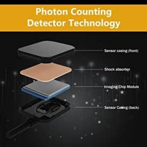
Photon Counting Device (PCD) Technology in Dental Radiology Sensors
Photon Counting Device (PCD) based sensors represent a cutting-edge development in dental radiology.
PCD technology works by directly counting individual X-ray photons and converting them into digital signals. This results in higher precision and sensitivity compared to other sensor technologies.
- Key advantages of PCD technology include:
- Superior image quality with high contrast resolution
- Lower radiation dose requirements
- Reduction of noise and artifacts
PCD Technology: The Future of Dental Radiology
PCD technology offers significant advantages over CMOS and CCD sensors, making it a compelling choice for dental practices seeking state-of-the-art imaging solutions.
Some of the primary benefits of PCD technology include:
Improved diagnostic capabilities: PCD sensors deliver higher image quality and better contrast resolution, enabling dentists to diagnose oral health conditions more accurately and with greater confidence.
Enhanced patient safety: By requiring lower radiation doses, PCD technology reduces patients’ exposure to ionizing radiation, promoting a safer dental environment.
Reduced noise and artifacts: PCD technology’s direct photon counting approach minimizes image noise and artifacts, resulting in clearer and more reliable images.
Conclusion:
Understanding the differences between CMOS, CCD, and Photon Counting Device technology in dental radiology sensors is essential for dentists looking to invest in the most suitable imaging solution for their practice.
While CMOS and CCD sensors have their advantages, PCD technology offers superior image quality, lower radiation dose requirements, and reduced noise, making it a promising option for the future of dental radiology.


Leave a comment