
The Diagnostic Powerhouse: Elevating Dental Care with 3D Imaging
The landscape of dental care has undergone significant transformation over the years, with advancements in technology paving the way for more accurate diagnoses, efficient treatment plans, and improved patient outcomes. At the forefront of these advancements is the evolution of dental imaging technologies. From the early days of conventional two-dimensional X-rays to the latest in high-definition imaging, the journey of dental diagnostics has been marked by continual innovation and refinement.
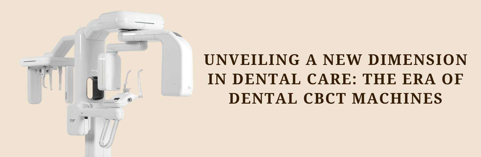
Enter the era of 3D dental diagnostic tools, among which Dental Cone Beam Computed Tomography (CBCT) Imaging stands out as a revolutionary advancement. Dental CBCT has transformed the way dental professionals approach diagnostics, offering a three-dimensional view of dental structures, nerves, and soft tissues with unprecedented clarity and detail. This leap in imaging capability allows for a more thorough analysis than ever before, making it an indispensable tool in modern dental practice.
The advent of Dental CBCT Imaging marks a significant milestone in dental diagnostics, providing a depth of insight that was previously unattainable with traditional radiography. Its ability to deliver precise, three-dimensional images has opened up new possibilities in treatment planning and disease management, making it a cornerstone of advanced dental care. As we delve deeper into the capabilities and applications of this powerful diagnostic tool, it’s clear that Dental CBCT Imaging is not just an enhancement of existing technologies but a paradigm shift in how dental health is assessed and treated.

Understanding Dental CBCT Imaging
Dental Cone Beam Computed Tomography (CBCT) represents a monumental leap forward from traditional dental radiography solutions. Unlike standard two-dimensional X-rays, CBCT technology offers three-dimensional imaging, granting dental professionals an unparalleled view of dental structures, soft tissues, nerve paths, and bone in the craniofacial region from a single scan.
CBCT functions by rotating around the patient, capturing hundreds of images from different angles. These images are then reconstructed to produce a 3D model of the patient’s dental anatomy. This method provides a more comprehensive and detailed view compared to the flat images produced by traditional X-rays, enabling a level of precision in dental imaging previously unattainable.
The technology behind CBCT is designed with the patient’s safety and comfort in mind, utilizing a lower dose of radiation compared to conventional computed tomography (CT) scans while delivering superior image quality. This aspect is crucial for recurrent diagnostics and follow-ups, ensuring patient exposure is kept to a minimum.
One of the unique advantages of CBCT is its ability to support a wide range of dental diagnostic requirements, from implant planning and orthodontic assessment to the evaluation of complex dental and maxillofacial conditions. Its precision imaging significantly enhances the accuracy of diagnostics, treatment planning, and surgical predictability, making it an indispensable tool in modern dentistry.
In essence, Dental CBCT imaging has transformed dental diagnostics, offering depth, clarity, and a comprehensive perspective that far surpasses traditional radiography. Its adoption into dental practices marks a significant advancement in how professionals’ approach, plan, and execute dental care, setting a new standard in precision dental imaging.

The Versatility of CBCT in Dental Care
Dental Cone Beam Computed Tomography (CBCT) imaging technology has revolutionized dental care, offering unparalleled precision and versatility across a myriad of dental specialties. Its applications range from implant planning and orthodontics to complex maxillofacial analysis, each benefiting from the detailed insights that CBCT provides.
Implant Planning CBCT
In the realm of dental implants, precision is paramount. CBCT imaging plays a crucial role in enhancing the accuracy and safety of implant procedures. By providing a 3D view of the patient’s bone structure, CBCT allows for meticulous pre-surgical planning. Dental professionals can assess bone quality and density, identify the optimal placement for implants, and avoid critical structures such as nerves and sinuses. This level of detail ensures a higher success rate for implants, with reduced risk of complications.
Orthodontic Diagnostic Imaging
For orthodontic purposes, CBCT imaging facilitates a comprehensive assessment of the dentofacial structure, offering clear insights into tooth orientation, jaw relationships, and bone anomalies. This detailed assessment aids in crafting more effective and individualized treatment plans. CBCT has become indispensable in orthodontics, particularly for complex cases where traditional imaging might not reveal the full picture, enabling practitioners to predict treatment outcomes with greater confidence.
Maxillofacial CBCT Analysis
CBCT imaging excels in providing critical insights into complex cases involving the jaw, facial structures, and sinuses. Whether it’s assessing the impact of trauma, diagnosing pathologies, or planning surgical interventions, CBCT offers a detailed view of the maxillofacial region. This capability is invaluable for surgeons and specialists who require a comprehensive understanding of anatomical relationships and pathologies to make informed decisions regarding treatment and surgical approaches.
The versatility of CBCT in dental care underscores its status as a diagnostic powerhouse. By offering detailed, three-dimensional images across various dental specialties, CBCT technology has significantly improved the way dental professionals diagnose, plan, and execute treatments, contributing to better patient outcomes and advancing the field of dental care.
CBCT for Dental Professionals: Benefits and Considerations
The adoption of Cone Beam Computed Tomography (CBCT) in dental practices offers a myriad of benefits that significantly enhance the quality-of-care dental professionals can provide. However, alongside these advantages, certain considerations must be taken into account to ensure the optimal use of this technology.
Benefits of CBCT for Dental Professionals
Improved Diagnostic Accuracy: One of the most significant benefits of CBCT is its ability to provide highly accurate and detailed 3D images. This accuracy is crucial for diagnosing complex dental issues, allowing for a level of detail that surpasses traditional 2D imaging techniques.
Comprehensive View of Dental Structures: CBCT offers a complete and comprehensive view of the patient’s dental anatomy, including bones, teeth, nerve pathways, and soft tissues. This all-encompassing perspective is invaluable for planning treatments across various dental specialties, from orthodontics to oral surgery.
Enhanced Treatment Planning: With the detailed imagery provided by CBCT, dental professionals can plan treatments with greater precision. This includes everything from the placement of dental implants to the strategic movement of teeth in orthodontic procedures, ensuring optimal outcomes for patients.
Considerations When Using CBCT
Radiation Exposure: While CBCT uses a lower dose of radiation compared to traditional medical CT scans, it’s still higher than standard dental X-rays. Dental professionals need to consider the risk-benefit ratio for each patient, reserving CBCT scans for cases where the detailed information it provides is essential for diagnosis or treatment planning.
Importance of Proper Training for Interpretation: The detailed images produced by CBCT require a trained eye for accurate interpretation. Dental professionals must undergo proper training to effectively analyze CBCT scans, ensuring that diagnoses are accurate and that the full benefits of this technology are realized.
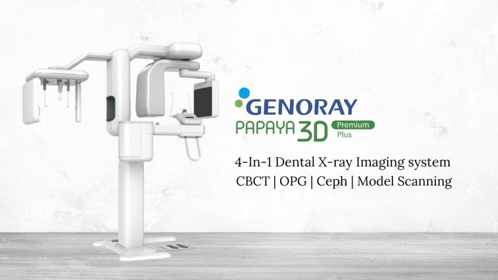
Highlighting Genoray Papaya 3D Plus
Among the CBCT systems available, the Genoray Papaya 3D Plus stands out for its exceptional image quality and user-friendly interface. Designed with dental professionals in mind, it provides a seamless experience from scan to diagnosis, equipped with advanced features that facilitate accurate implant planning, orthodontic assessments, and comprehensive maxillofacial analysis. Its inclusion in a dental practice represents a commitment to leveraging state-of-the-art technology for enhanced patient care.
In summary, the introduction of CBCT, exemplified by systems like the Genoray Papaya 3D Plus, has transformed dental diagnostics and treatment planning. While the benefits are substantial, dental professionals must also consider the implications of radiation exposure and the necessity of proper training to fully harness the potential of this advanced imaging technology.
Patient Outcomes and CBCT: A Closer Look
The introduction of Cone Beam Computed Tomography (CBCT) into dental practice has not only revolutionized diagnostic and treatment planning processes but has also significantly improved patient outcomes. The capability of CBCT to provide detailed 3D images leads to more precise diagnoses, the possibility of minimally invasive treatment options, and the creation of highly personalized care plans.
Precise Diagnosis
CBCT imaging’s precision plays a pivotal role in diagnosing various dental conditions, from identifying the early stages of periodontal disease to detecting complex root canal anatomy. This level of detail ensures that conditions are not just identified but thoroughly understood, allowing for targeted treatments that address the root cause of the issue, rather than just the symptoms. As a result, patients receive care that is not only effective but also efficient, reducing the need for multiple treatments or corrections down the line.
Minimally Invasive Treatment Options
With the detailed view provided by CBCT scans, dental professionals can plan and execute treatments that are less invasive than traditional methods. For example, in implant dentistry, CBCT imaging allows for the precise placement of implants without extensive cutting and suturing of gum tissue. This precision reduces patient discomfort and speeds up the recovery process, making dental procedures a less daunting prospect for patients.
Personalized Care Plans
CBCT technology enables the creation of personalized treatment plans tailored to the individual’s specific anatomy and needs. This customization is particularly beneficial in orthodontics, where CBCT imaging helps in planning tooth movements and orthodontic interventions with a level of accuracy previously unattainable.
Case Studies Illustrating CBCT’s Impact
– Implant Planning: A patient presented with a missing molar and limited bone density, complicating implant placement. A CBCT scan allowed the dentist to visualize the bone structure in 3D, identify a suitable location for the implant, and plan the surgery with minimal risk. The implant was placed successfully, with optimal osseointegration and patient satisfaction.
– Orthodontic Assessment: In another case, a teenager with complex orthodontic issues, including impacted teeth and jaw misalignment, underwent CBCT scanning. The detailed imagery provided a clear roadmap for treatment, enabling the orthodontist to devise a strategy that addressed all issues in a coordinated manner. The result was a more efficient treatment course with outstanding aesthetic and functional outcomes.
The Future of Dental Diagnostics: Trends and Innovations
The landscape of dental diagnostics is poised for transformation, driven by rapid advancements in technology and a deeper understanding of oral health’s complexities. As 3D dental diagnostic tools and imaging technologies continue to evolve, the future promises even greater precision, efficiency, and personalized care in dentistry. Two of the most exciting frontiers in this evolution are the integration of artificial intelligence (AI) and machine learning (ML) into dental imaging.
Advancements in 3D Dental Diagnostic Tools
Future advancements in 3D dental diagnostic tools are expected to offer even higher resolution images, with faster scanning times and more user-friendly interfaces. These improvements will further reduce the need for retakes, minimize patient discomfort, and enhance diagnostic accuracy. Additionally, portable and more compact CBCT scanners could become more prevalent, making advanced imaging technology accessible to a broader range of dental practices, including those in rural or underserved areas.
Integration of AI and Machine Learning
The integration of AI and machine learning into dental imaging represents a groundbreaking shift. AI algorithms are being developed to analyze 3D images with remarkable speed and accuracy, identifying patterns and anomalies that might escape the human eye. This capability could significantly enhance the detection of early-stage diseases, predict treatment outcomes, and suggest optimal treatment plans based on vast datasets of dental cases.
Machine learning, a subset of AI, could further refine the diagnostic process by learning from each scan it analyzes, continuously improving its diagnostic capabilities. This could lead to more accurate assessments of complex conditions, such as predicting the progression of periodontal disease or assessing the risk of complications from dental surgeries.
Enhanced Image Analysis and Interpretation
AI and machine learning will not only automate image analysis but also offer more detailed interpretations, providing dentists with insights into patient health that were previously unavailable. This could include the automatic measurement of bone density, the detection of micro-fractures, and even the assessment of the risk of future dental problems.
Personalized Treatment Plans
The future of dental diagnostics will likely also see a more personalized approach to treatment planning. With AI-driven analysis, dentists could receive recommendations for the most effective treatment options tailored to the individual patient’s specific condition, anatomy, and health history, further improving patient outcomes.
Conclusion: The Role of 3D Imaging in Transforming Dental Practice
The advent and integration of Cone Beam Computed Tomography (CBCT) and 3D imaging technologies into dental practice have marked a significant turning point in the provision of dental care. These advanced diagnostic tools have fundamentally transformed the landscape of dentistry, elevating the standard of care to unprecedented levels. By offering detailed, three-dimensional views of the oral and maxillofacial regions, CBCT and 3D imaging have enhanced diagnostic accuracy, enabled more precise treatment planning, and opened the door to minimally invasive treatment options.
The comprehensive insights provided by these technologies allow dental professionals to approach each case with a depth of understanding previously unattainable. From implant planning and orthodontic assessment to complex maxillofacial surgery, the applications of CBCT and 3D imaging are vast, underscoring their pivotal role in modern dentistry. These tools have not only improved the outcomes of dental treatments but have also significantly enhanced patient experiences, reducing discomfort and treatment times while increasing satisfaction.
As we look to the future, the potential for further advancements in dental diagnostic technologies is vast. The integration of artificial intelligence and machine learning promises to unlock new capabilities in image analysis and interpretation, further refining the precision and personalization of dental care.
In light of these advancements, it is incumbent upon dental professionals to embrace and integrate these technologies into their practices. Staying abreast of the latest developments in 3D imaging and diagnostic tools is essential for maintaining a competitive edge in the field and, more importantly, for continuing to improve the quality of care provided to patients.

Leave a comment